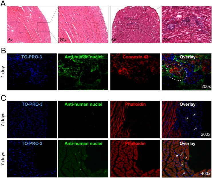
Fig 8. In vivo transplantation of spheroids in myocardium-injured mice.
(A) Evaluation of cardiotoxin-induced injury in the myocardium wall at two magnifications (right) compared to healthy myocardium (left) in Hematoxylin-Eosin staining. (B) Myocardium sections from myocardium injured mice transplanted with spheroids were stained with TO-PRO3 (blue) to show all nuclei, anti-human nuclei antibody (green), and connexin-43 or phalloidin (red) for actin and analysed by confocal microscopy. Representative experiments out of three performed are shown. The circle and arrows show the engrafted spheroid 1 day after the injection and the dispersed hCPCs 7 days after the injection, respectively.

