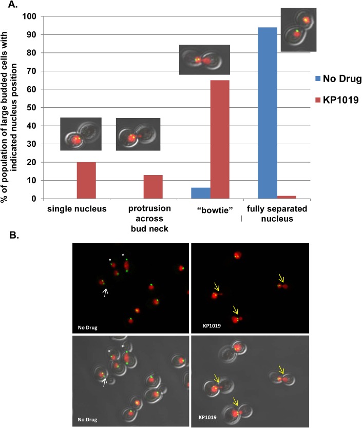
Fig 5. Nuclear Morphology in KP1019 treated cells.
A. Early log phase wild-type yeast were treated with 80ug/ml KP1019 for three hours and nuclear morphology was determined as described in Materials and Methods. Large budded cells were scored on the position of the nucleus relative to the bud neck. Categories are indicated on the X-axis, and include a single nuclei positioned near the bud neck and not migrating across the bud neck (single nucleus), cells with some protrusion of nucleus across bud neck (protrusion across bud neck), nuclei spanning the bud neck (bowtie), and two separated nuclei positioned in each cell body (separated). Example image of treated cells from each category is provided. B. Representative images from the cytological analysis used to obtain data in Parts A showing varying levels of nuclear migration across the bud neck. Red signal is Histone H2-mCherry indicating nuclear position. Green signal is Spc42-GFP indicating SPB position. Arrows indicate cells with short spindles; the white color indicates an untreated cell with a single nuclei, and the yellow color indicates treated cells showing migration of nuclei into the daughter cell. White asterisk show fully separated spindle in untreated cells at varying points in anaphase. Note more random orientation of SPB bodies relative to the longitudinal cell axis exampled in treated cells (S1 Fig).

