
Figure 3.
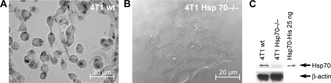
Target specificity of antibody-coated gold nanoparticle (Au-Hsp70) uptake.
Notes: 4T1 mouse mammary carcinoma cells were incubated with antibody-coated nanoparticles (1 µg/mL) for 24 hours at 37°C. (A) In 100% of the 4T1 cells with a wild-type expression of Hsp70, cmHsp70.1-coated nanoparticles could be detected as black dots (arrows). (B) In transfected cells, 4T1 Hsp70−/− cells with knocked down Hsp70, no uptake of nanoparticles could be detected. (C) The successful knock down of Hsp70 in 4T1 Hsp70−/− cells was verified by Western blotting compared with the expression of Hsp70 in the cytosol of 4T1 wt cells. Images are representative for at least three independent experiments. (A and B bright field, objective 40×, scale bar 20 µm.)
Abbreviation: wt, wild-type.