
Figure 1.
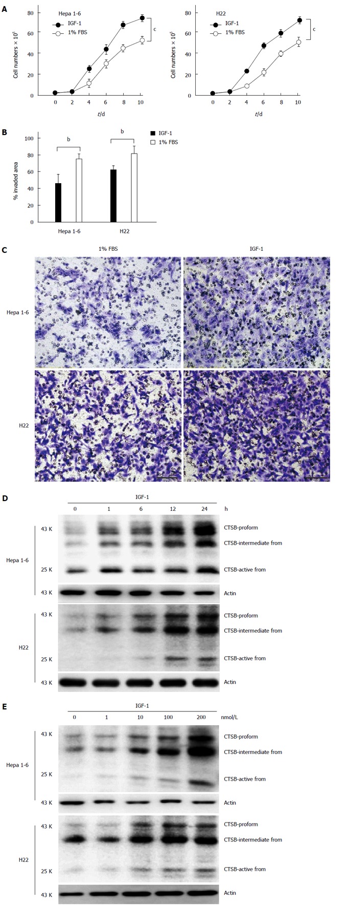
Cathepsin B mediates the IGF-1-induced hepatocellular carcinoma-promoting effect. A: Hepa 1-6 and H22 cells were treated with IGF-1 (100 nmol/L) for 10 d. The data are presented as representative growth curves; B: Wound healing assay of Hepa 1-6 and H22 cells treated with IGF-1 for 24 h; C: IGF-1 induced Hepa 1-6 and H22 cell invasion. In total, 1 × 10⁵ Hepa 1-6 and H22 cells that had been treated with IGF-1 for 24 h were allowed to invade through Transwell inserts (8 μm) coated with Matrigel. The cells on the lower surface of the chambers were stained and imaged. The data are presented as a representative Transwell assay (scale bar, 20 μm); D and E: Hepa 1-6 and H22 cells were treated with IGF-1 (100 nmol/L) for the indicated times or treated with different IGF-1 concentrations for 12 h. CTSB expression was determined by immunoblotting. The data are presented as representative immunoblots. Student’s t-test. bP < 0.01; cP < 0.001, IGF-1 group vs 1% FBS group.