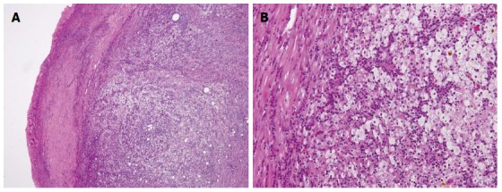
Figure 7.

Microscopic examination revealing a gallbladder wall markedly thickened with severe inflammation and fibrosis (A), HE × 200; Large xanthoma cell with clear-to-foamy lipid-containing cytoplasm and interspersed lymphocytes invading the liver (B), HE × 400.
