Figure 4.
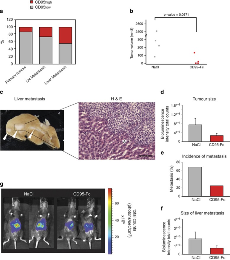
Blocking of CD95L reduces tumour size and metastasis of PDAC. (a) Semiquantitative analysis of CD95 immunostaining in TMA containing PDAC samples, and lymph node and liver metastases from different primary PDAC tumours. (b) Mice were orthotopically transplanted with PanD24 cells and at days 3 and 7 after transplantation animals were treated with 50 μg CD95-Fc i.p. After 105 days, mice were killed and tumour volumes were assessed with a caliper. Treatment with CD95-Fc (n=4) clearly reduced tumour volumes compared with the NaCl- (n=4) treated control group (Wilcoxon's rank-sum test, P-value=0.05714). (c) Picture of a liver from a control mouse with macroscopic liver metastasis, indicated by white arrows (left side), and a representative picture of hematoxylin and eosin (H&E)-stained liver metastasis (right side). No metastatic lesions were detected in the livers of mice treated with CD95-Fc. Asterisk denotes liver tissue. Scale bar: 20 μm. (d–g) Mice were orthotopically injected with Panc02 cells and at days 3 and 7 after injection animals were treated with 50 μg CD95-Fc (n=16) or NaCl (n=14) intravenously. Three weeks later, mice were killed and tumour size (d) and liver metastases (f) was assessed by bioluminescence imaging. (e) Percentage of animals with detectable liver metastases. (g) Representative bioluminescence pictures. Results are expressed as mean±S.E.M.

