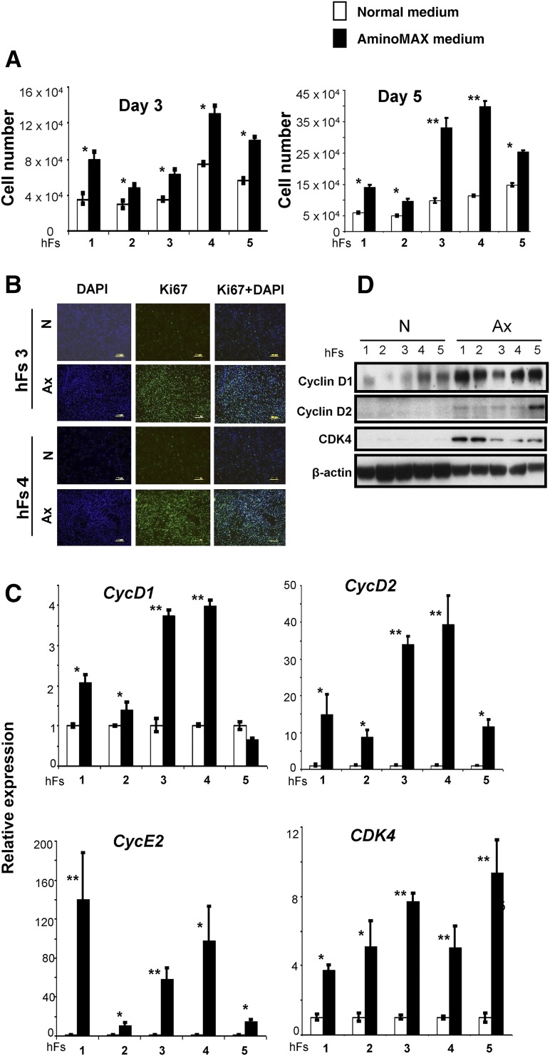
Figure 1.
The growth medium determines growth kinetics of human somatic fibroblasts. (A): The cell numbers of 5 primary hFs—AG16104 (hFs 1), AG16086 (hFs 2), 120111 (hFs 3), 120116 (hFs 4), and AG16102 (hFs 5)—were measured by a hemocytometer on day 3 and day 5 after excluding dead cells by trypan blue staining. (White boxes represent cell numbers in N and black boxes indicate cell numbers in defined Ax.) The cells were seeded at equal densities (30,000 cells per well) on day 0 and counted on day 3 and day 5 in triplicate wells. The x-axis denotes the hFs. (B): hFs 3 and hFs 4 were seeded at equal densities (30,000 cells per well) and cultured in N or Ax medium. Cells were harvested on day 3 and subjected to Ki67 immunostaining to identify cycling cells. DAPI was used to stain nuclei. Scale bars = 200 μm. (C): The relative expression of cell cycle genes CycD1, CycD2, CDK4, and CycE2 in hFs grown in N (white bars) or Ax medium (black bars). Expression was normalized to the β-actin gene and is shown relative to the average N medium level. The x-axis denotes the number of hFs. (D): Expression of CycD1, CycD2, and CDK4 proteins was demonstrated by Western immunoblot analysis. β-actin was used as an internal control. All experiments were performed three times. Data are shown as mean ± SD. Statistical significance was determined by Student’s t test. ∗, p < .05; ∗∗, p < .01 for Ax versus N. Abbreviations: Ax, AminoMAX medium; DAPI, 4,6-diamidino-2-phenylindole; hF, human fibroblast; N, Dulbecco’s Modified Eagle’s medium.

