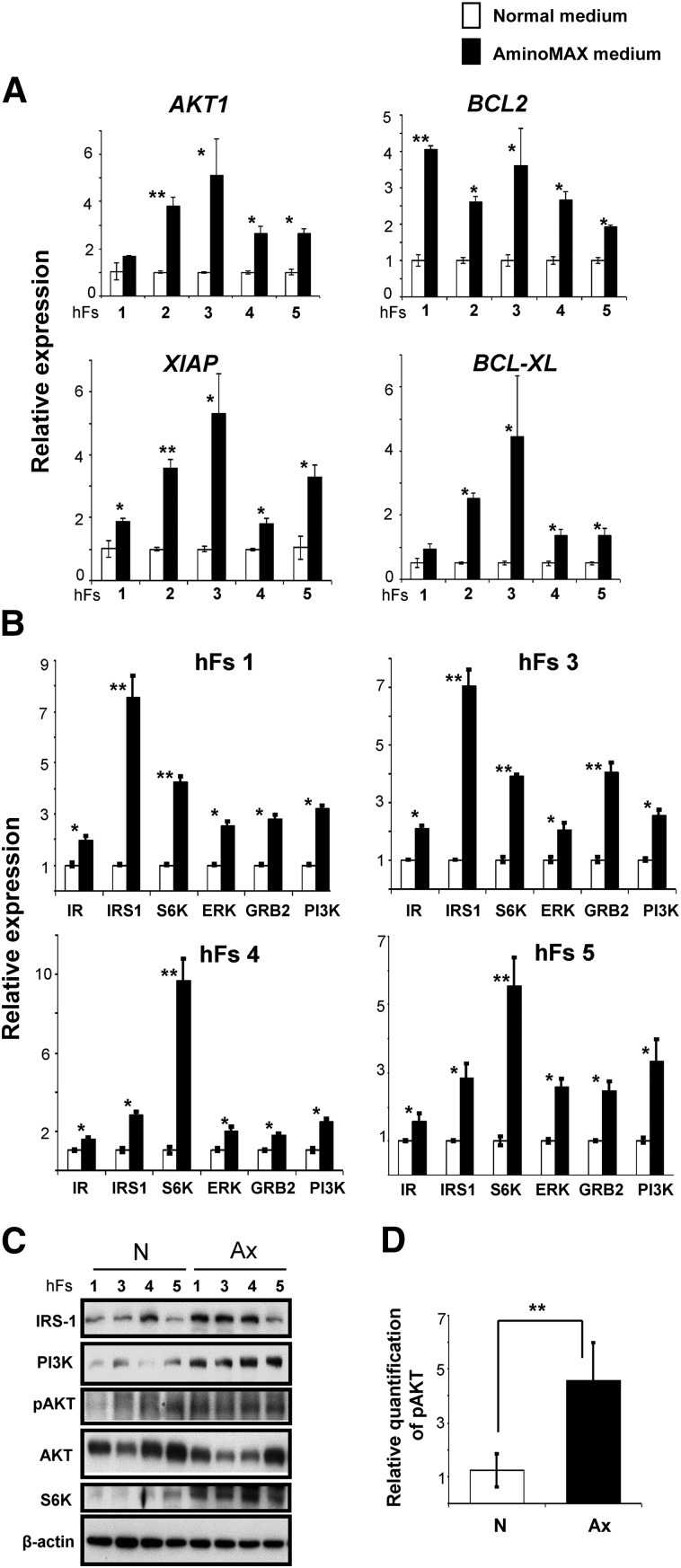
Figure 2.
Fibroblasts cultured in Ax growth medium exhibit altered expression of genes in cell survival and growth factor (insulin/insulin-like growth factor-1 [IGF-1]) signaling pathways. (A): The relative expression of AKT1, BCL2, XIAP, and BCL-XL genes by real-time polymerase chain reaction in human fibroblasts (hFs 1, 2, 3, 4, and 5) cultured in N (white bars) or Ax (black bars) medium. The x-axis denotes the hFs. (B): Gene expression analysis of proteins in the growth factor (insulin/IGF-1) signaling pathway, including IR, IRS1, S6K, ERK, GRB2, and PI3K, in four hFs (hFs 1, hFs 3, hFs 4, and hFs 5) grown in N or Ax medium. In both (A) and (B), expression was normalized to the β-actin gene and is shown relative to the average N medium level. The x-axis denotes the number of hFs. (C): Western blot analysis of IRS1, PI3K, S6K, pAKT, and AKT proteins in hFs 1, hFs 3, hFs 4, and hFs 5 cultured in N or Ax medium. β-Actin was used as an internal control. (D): Graph representing the relative quantity of phosphorylation of AKT normalized to total AKT band density by ImageJ software (US National Institutes of Health, Bethesda, MD, http://imagej.nih.gov/ij). All experiments were performed three times, represented as mean ± SD. Statistical significance was determined by Student’s t test. ∗, p < .05; ∗∗, p < .01 for Ax versus N. Abbreviations: Ax, AminoMAX medium; D, day; ERK, extracellular signal-regulated kinase; GRB2, growth factor receptor-bound protein; IR, insulin receptor; IRS1, insulin receptor substrate-1; N, Dulbecco’s modified Eagle’s medium; PI3K, phosphatidylinositol 3-kinase; S6K, S6 kinase.

