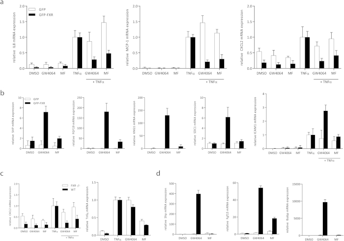
Figure 3. MF reduces pro-inflammatory gene expression in HepG2 cells and intestinal organoids.
Endogenous FXR target gene expression in HepG2 cells stably overexpressing GFP (HepG2-GFP; white bars) or GFP-FXR (HepG2-GFP-FXR; black bars). (A) Cells were treated in triplicate with DMSO, 1 μM GW4064, 10 μM MF, 5 ng/ml TNFα, GW4064 plus TNFα, or TNFα plus MF for 24 hours. IL8, MCP-1, and CXCL2 mRNA expression was analyzed by qRT-PCR in duplicate. (B) HepG2-GFP and HepG2-GFP-FXR cells were treated with DMSO, 1 μM GW4064 or 10 μM MF in triplicate for 24 hours. SHP, FGF19, KNG1, SDC1 and ICAM1 mRNA expression was analyzed by qRT-PCR in duplicate. Each bar represents mean ± SD. (C,D) Small intestine derived organoids from 3WT and FXR−/− mice were treated with DMSO, 1 μM GW4064, 10 μM MF, 5 ng/ml TNFα, GW4064 plus TNFα, or TNFα plus MF for 24 hours. mRNA expression of each organoid line was analyzed by qRT-PCR in duplicate. Each bar represents mean ± SEM.

