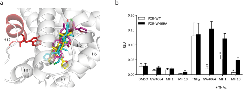
Figure 4. Superposition of the three predicted MF binding modes.

Three different binding modes of MF to FXR, as determined by docking studies, are depicted. (A) Binding mode 1 suggests that the furoate group of MF is buried into the FXR binding site pointing towards helix 7 (yellow carbons); binding mode 2 is characterized by the furoate group of MF oriented toward helix 11 and helix 12 (blue carbons); binding mode 3 is head-to-tail flipped with respect to binding modes 1 and 2 with the furoate group of MF oriented toward the helix 5 and 6 (magenta carbons). Helix 12 is shown in red. (B) HEK293T cells transfected with NF-κB reporter, expression plasmids for wild type FXR (FXR-WT) or mutant FXR (FXR-W469A) and RXR, and pTK-renilla construct were treated with DMSO, 1 μM GW4064, 1 or 10 μM MF as indicated, or TNFα, for 24 hours. The NF-κB luciferase reporter assay was performed in quadruplicate. *p < 0.001 compared to FXR-W469A cells treated with GW4064 or MF plus TNFα.
