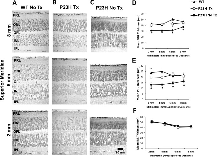
Figure 1.
Representative photomicrographs of the retina taken along the superior meridian 2, 6, and 8 mm above the optic disc (A–C). WT (No Tx N = 6; WT Tx N = 2; column A) and Tg P23H Curcumin-Tx (N = 4; column B) sections appeared similar and showed the normal laminar arrangement of the retina. In contrast, Tg P23H No Tx (N = 8; column C) showed obvious thinning of the ONL. Morphometric analysis of the ONL, PRL, and INL (D–F). (D) Tg P23H No Tx miniswine embryos showed a central-to-peripheral thinning of the ONL that was significantly reduced (black asterisks) in several retinal locations compared with WT and Tg P23H Tx, which were similar. The mean thickness in the WT showed a significant increase at 6-mm superior to the disc, whereas the treated Tg P23H Tx remained the same thickness from 2- to 8-mm superior to the disc (gray asterisk). (E) P23H No Tx retinas showed a significant central-to-peripheral thinning of the PRL, while PRL thickness was similar in WT and Tg P23H Tx miniswine embryos. (F) Quantitative measurements of the INL appeared similar across all groups and retinal locations. Scale bar: 20 μm and applies to columns A to C.

