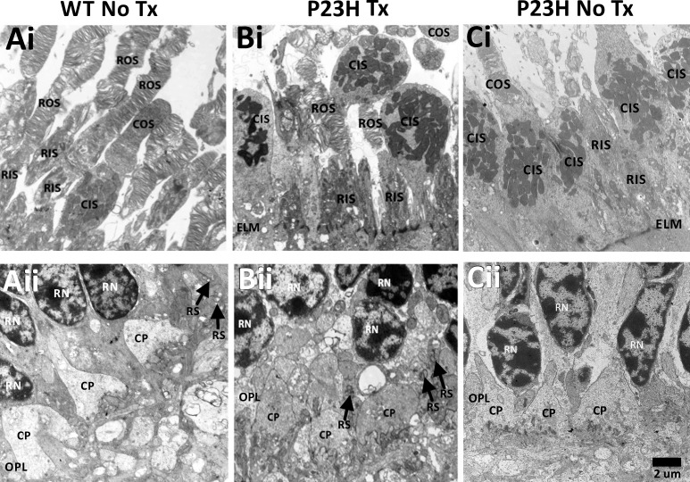
Figure 2.
Transmission electron micrographs of the PRL (Ai–Ci) and OPL (Aii–Cii). WT (Ai) and P23H Tx (Bi) showed ROS and RIS, and COS and CIS, while P23H No Tx (Ci) lacked identifiable ROS, but did have CIS and COS. The ELM appeared intact in all images. WT (Aii) and P23H Tx (Bi-i) showed RS with triadic profiles (black arrows) and CP in the OPL, while untreated P23H (Cii) lacked RS, but did have CP in the OPL. CIS, cone inner segments; COS, cone outer segments; CP, cone pedicles; ELM, external limiting membrane; OPL, outer plexiform layer; RIS, rod inner segments; RN, rod nuclei; ROS, rod outer segments; RPE, retinal pigment epithelium; RS, rod spherules. Scale bar: 2 μm and applies to all panels.

