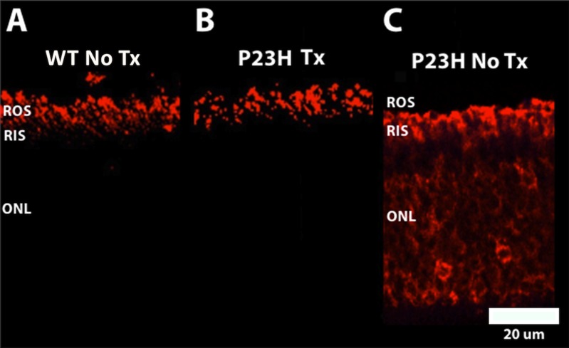
Figure 3.
Representative retinal micrographs of rhodopsin localization detected by immunolabeling with anti-Rho 1D4 (rhodopsin) antibody. WT (A) and P23H Tx (B) showed normal localization of rhodopsin (red labeling) that was confined to the ROS. In contrast, P23H No Tx (C) showed mislocalization of rhodopsin to the RIS and ONL (red labeling). Scale bar: 20 μm and applies to all panels.

