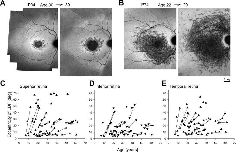
Figure 2.
Long-term progression of the LDF as determined from wide-angle NIR-RAFI performed serially in ABCA4-RD along the superior, inferior, and temporal meridians. (A) Representative progression in a patient (P34) with a central macular lesion; NIR-RAFI recorded over a 9-year interval at ages 30 and 39. (B) Representative progression in a patient (P74) with a lesion extending well beyond the macula; NIR-RAFI recorded over a 7-year interval at ages 22 and 29. (C–E) Eccentricity of the LDF in ABCA4-RD as a function of age along the superior (C), inferior (D), and temporal (E) meridians. Symbols connected by lines represent serial follow-up in the same eye at two visits.

