
Figure 2.
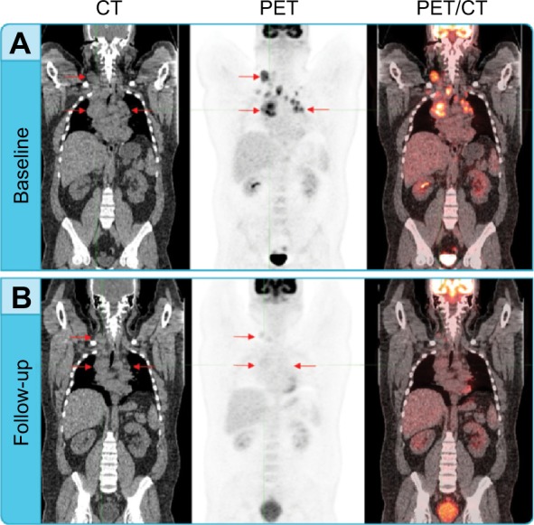
Assessing treatment response using PET and CT.
Notes: (A) Baseline coronal CT, PET, and fused PET/CT images of a patient with lymphoma. Multi-focal bilateral FDG-avid adenopathy, including a large right superior mediastinal mass lesion (arrows) with marked focal FDG uptake visible on the coregistered FDG-PET consistent with lymphoma. (B) Follow-up coronal CT, PET, and PET/CT images of the same patient after two cycles of therapy. The multi-focal adenopathy including the large right superior mediastinal mass is still visible on the CT image and the right mediastinal lesion appears stable (arrows). The PET image demonstrates complete resolution of tumor metabolic activity. All previous FDG-avid regions are indiscernible from background, consistent with a CMR. The CMR noted on the PET examination indicates a treatment response before any change is visible by CT.
Abbreviations: CMR, complete metabolic response; CT, computed tomography; FDG, fluorodeoxyglucose; PET, positron emission tomography.