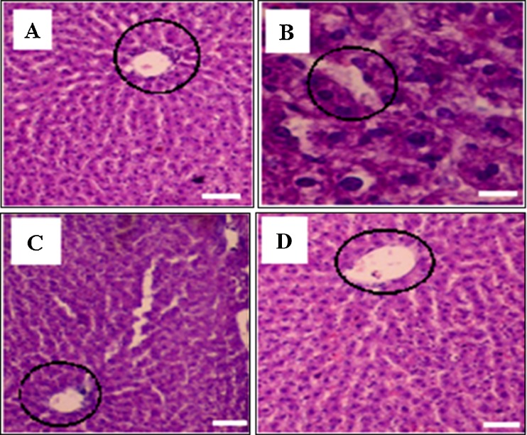
Fig. 4.
Histopathological observation of liver of different treated groups (60x). Section through the liver of normal rats showing central vein and hepatocytes (a), section through the liver of CCl4-treated rats showing central vein and hepatocytes (b) and section through the liver of ASX, ASXEs (250 μg/kg b.w) treated rats (c and d) showing the central vein (round marking) and hepatocytes

