Fig. 1.

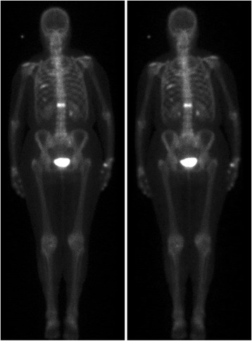
Images corresponding to a typical patient. Left: image acquired using the standard full-time protocol. Right: image acquired using the half-time protocol and processed with the Pixon-algorithm

Images corresponding to a typical patient. Left: image acquired using the standard full-time protocol. Right: image acquired using the half-time protocol and processed with the Pixon-algorithm