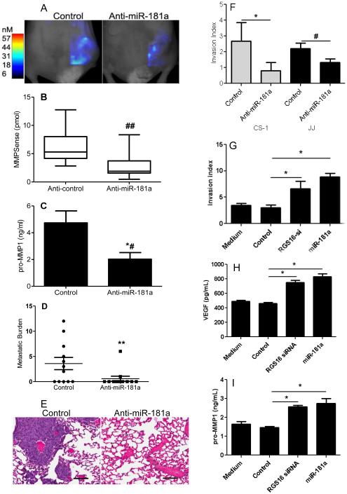
Figure 4. Anti-miR181a inhibits chondrosarcoma invasion and metastasis.
CS-1 cells expressing either control or anti-miR181a were used for in vitro invasion assay and for xenograft tumors as described in Methods. After 3 weeks, xenograft tumors were evaluated with Fluorescence-based Tomography using the MMPSense probe and after 5 weeks lung metastatic burden was determined. (A) Representative imaging with MMPsense probe is shown. (B) Summary of imaging data with MMPSense probe, graphs represent the median, box represents 25th-75th percentiles, bars represent range. N=12/group, ## p<0.004. (C) MMP1 content in xenograft tumors was measured by ELISA, N=8/group, *# p <0.02. (D) metastatic burden was quantified as the number of lung sections per mouse with metastases. N=12 mice/group, ** p<0.03. (E) H&E stained lung demonstrating metastases. (F) In vitro invasion index results are shown for CS-1 and JJ cells (* p<0.001, N=9; # p<0.01) N=4/group). CS-1 cells were transfected with either control siRNA and control miR (control), siRNA RGS16, or miR-181a. Results of invasion assay (G), and ELISA for VEGF (H) and MMP1 (I) in conditioned media are shown. * p < 0.001, N = 4.

