Figure 6.

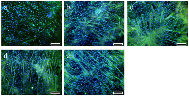
Tubulin (green) and nuclei (blue) staining of C2C12 cells after 7 days in differentiation medium on HPLA and HPLAAT substrates: (a) HPLA, (b) HPLAAT3, (c) HPLAAT6, (d) HPLAAT9 and (e) HPLAAT12. Scale bar: 200 μm.

Tubulin (green) and nuclei (blue) staining of C2C12 cells after 7 days in differentiation medium on HPLA and HPLAAT substrates: (a) HPLA, (b) HPLAAT3, (c) HPLAAT6, (d) HPLAAT9 and (e) HPLAAT12. Scale bar: 200 μm.