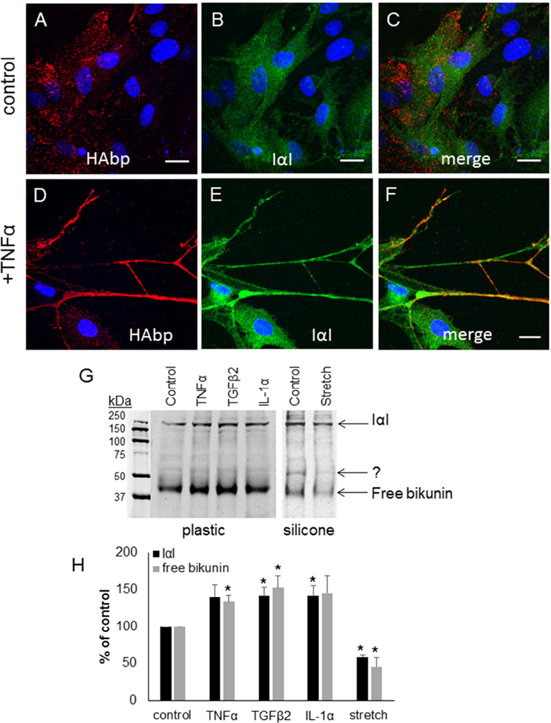
Figure 4. Bikunin and Inter-α-Inhibitor in TM cells.
Inter-α-inhibitor (green) was colocalized with HAbp (red) in control TM cells (A–C) or in TM cells treated for 3 days with TNFα (D–F). Merged images are shown (C, F). Confocal acquisition settings were maintained between untreated and TNFα-treated TM cells. DAPI was used to stain the nuclei (blue). Scale bars = 20 µm. (G) Western immunoblots of bikunin in media from untreated cells (control) and following treatment for 48 hours with TNFα, TGFβ2, IL-1α and mechanical stretch. The positions of free bikunin (42 kDa) and bikunin complexed with HCs to form IαI (240 kDa) are indicated. Equal amounts of protein were loaded into each lane. The cells on the left panel were cultured on plastic, whereas the cells in the right panel were cultured on collagen-coated silicone membrane. (H) Densitometry of bikunin bands are presented as a percentage of untreated control cells. N=3.* p < 0.05 by ANOVA.

