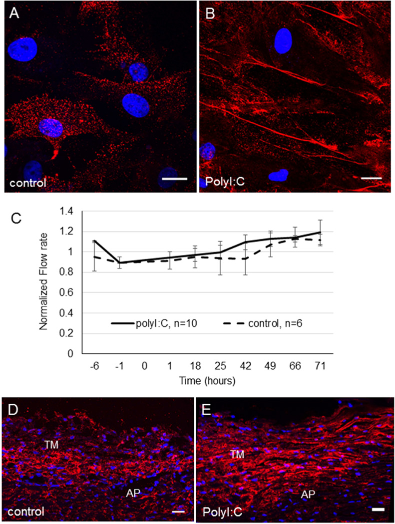
Figure 6. Induction of HA cables by polyI:C and the effect of polyI:C on outflow rates in porcine anterior segment perfusion culture.
TM cells in culture were (A) untreated (control) or (B) treated with 10 µg/ml polyI:C in PBS for 24 hours. HAbp was used to detect HA cable formation. DAPI was used to stain the nuclei (blue). Scale bars = 20 µm. (C) Porcine anterior segments were perfused with serum-free media. At time point 0, 10 µg/ml polyI:C in PBS was applied. Control anterior segments were untreated. Data shows the average flow rate ± standard error of the mean. N=10 for polyI:C; n=6 for control. HAbp staining of control porcine TM tissue (D) and polyI:C-treated TM (E). The cornea is positioned to the right in each image. AP = aqueous plexus. DAPI was used to stain the nuclei (blue). Scale bars = 20 µm.

