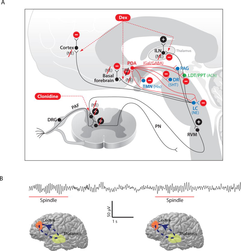
Fig. 10.

Neurophysiology of dexmedetomidine. A. Dexmedetomidine acts pre-synaptically to block the release of norepinephrine (NE) from neurons projecting from the locus coeruleus (LC) to the basal forebrain (BF), the pre-optic area (POA) of the hypothalamus, the intralaminar nucleus (ILN) of the thalamus and the cortex. Blocking the release of NE in the POA leads to activation of its inhibitory GABAergic (GABA) and galanergic (Gal) projections to dorsal raphé (DR) which releases serotonin (5HT), the tuberomamillary nucleus (TMN) which releases histamine (His), the LC, the ventral periacqueductal gray (PAG) which releases dopamine, the lateral dorsal tegmental (LDT) nucleus and the pedunculopontine tegmental (PPT) nucleus which release acetylcholine (Ach). These actions lead to decreased arousal by inhibition of the arousal centers. B. Ten-second electroencephalogram segment showing spindles, intermittent 9 to 15 Hz oscillations (underlined in red), characteristic of dexmedetomidine sedation. C. The spindles are most likely produced by intermittent oscillations between the cortex and thalamus (light green region). Panel A is reproduced with permission from Brown, Purdon and Van Dort, Annual Review of Neuroscience, 2011.
