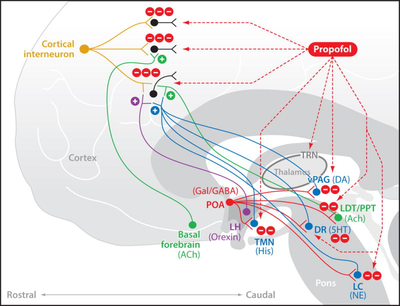
Fig. 4.

Neurophysiological mechanisms of propofol’s actions in the brain. Propofol enhances GABAA-mediated inhibition in the cortex, thalamus and brainstem. Shown are three major sites of action: post-synaptic connections between inhibitory interneurons and excitatory pyramidal neurons in the cortex; the GABAergic neurons in the thalamic reticular nucleus (TRN) of the thalamus; and post-synaptic connections between GABAergic projections from the pre-optic area (POA) of the hypothalamulus, and the monoaminergic nuclei which are the tuberomammillary nucleus (TMN) that releases histamine (His), the locus ceruleus (LC), that releases norepinephrine (NE), the dorsal raphé (DR) that releases serotonin (5HT); and the cholinergic nuclei which are the basal forebrain (BF), pedunculopontine tegmental (PPT) nucleus and the lateral dorsal tegmental (LDT) nucleus that release acetylcholine (Ach). This figure is reproduced with permission from Brown, Purdon and Van Dort, Annual Review of Neuroscience, 2011.
