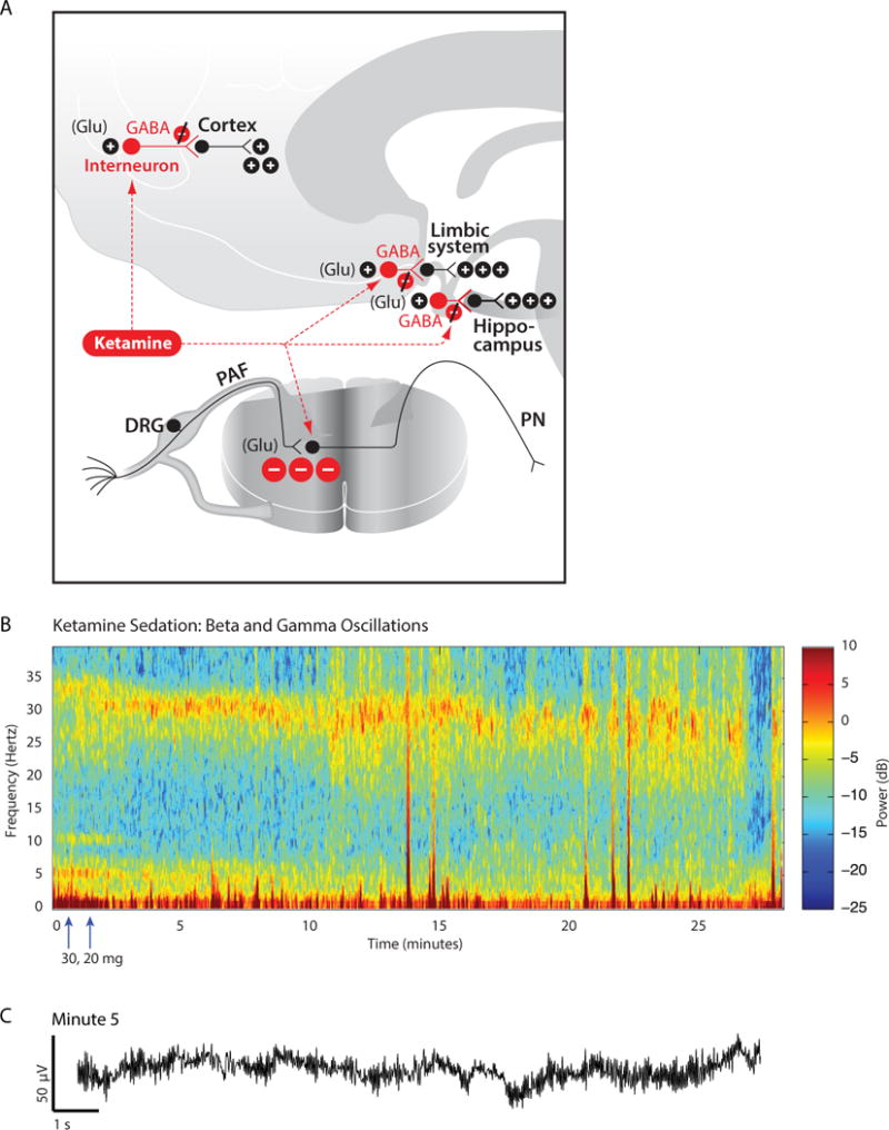
Fig. 9.

Neurophysiology and electroencephalogram signatures of ketamine. A. At low doses, ketamine blocks preferentially the actions of glutamate NMDA receptors on GABAergic inhibitory interneurons in the cortex and subcortical sites such as the hippocampus and the limbic system. The antinociceptive effect of ketamine is due in part to its blockade of glutamate release from peripheral afferent (PAF) neurons in the dorsal root ganglia (DRG) at their synapses on to projection neurons (PN) in the spinal cord. B. Spectrogram showing the beta-gamma oscillations in the electroencephalogram of a sixty-one year-old woman who received ketamine sedation for a vacuum dressing change. Blocking the inhibitory action of the interneurons in cortical and subcortical circuits helps explain why ketamine produces a high-frequency beta oscillation as its electroencephalogram signature. C. Ten-second electroencephalogram trace recorded at minute 5 from the spectrogram in B. Arrows indicates times of ketamine doses. Panel A is reproduced with permission from Brown, Lydic and Schiff, New England Journal of Medicine, 2010. Panels B and C were adapted from Purdon and Brown, Clinical Electroencephalography for the Anesthesiologist (2014), with permission from the Partners Healthcare Office of Continuing Professional Development.131
