
Fig. 4.
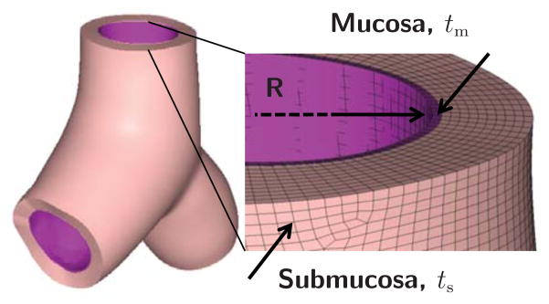
Representative Y-branch model created from magnetic resonance image segmentation. We segment the inner airway wall to generate a surface model, which we project outward to create a volume model parameterized in terms of the radius R and the mucosal and submucosal thickness offsets tm and ts. We mesh the volume model using linear hybrid hexahedral elements with four and ten elements across the mucosal and submucosal layers.