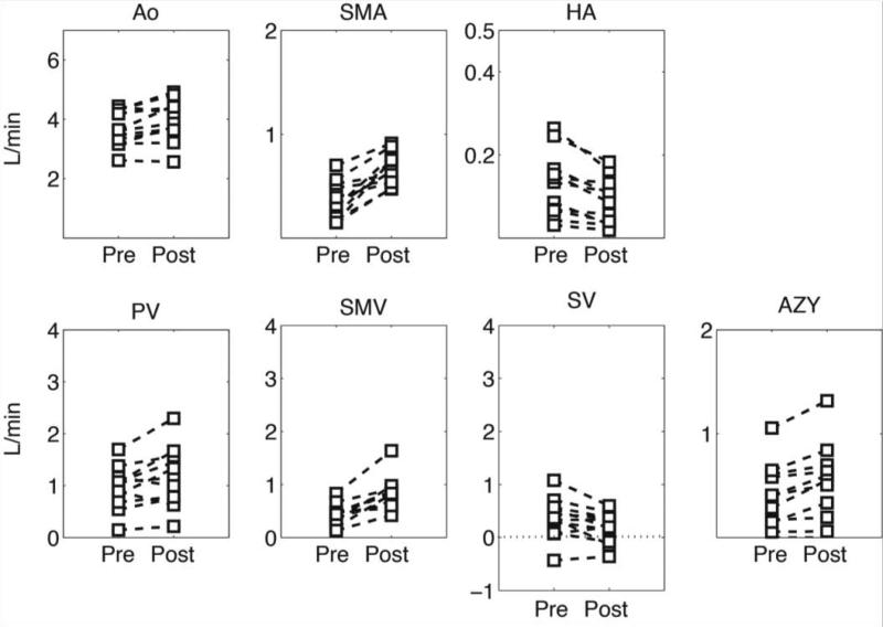
Figure 6.
Quantitative analysis in patients with cirrhosis. Blood flow was quantified at the supraceliac aorta (QAo), superior mesenteric artery (QSMA), hepatic artery (QHA), portal vein (QPV), superior mesenteric vein (QSMV), splenic vein (QSV) and azygos vein (Qazy). Note the hepatofugal flow (QSV) in some patients represented by the negative blood flow.

