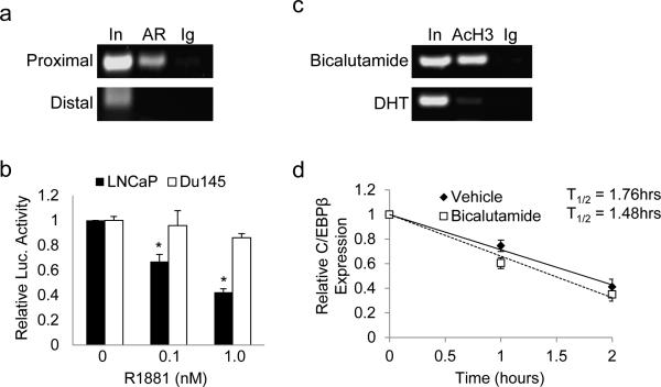
Figure 3.
AR binds and represses the CEBPB gene. (a) LNCaP cells were subjected to ChIP analysis with antibodies against androgen receptor (AR) or normal rabbit IgG (Ig). PCR was used to amplify DNA fragments centered at -187 bp (proximal) or an upstream fragment centered at -2040 bp (distal) of the CEBPB gene. (b) LNCaP or DU145 cells were transiently transfected with CEBPB-Luc and cultured with vehicle or R1881 at the indicated dose. Fold activation of the reporter relative to vehicle treated cells was determined after normalization to β-galactosidase activity. The average from 3 independent experiments is presented (c) LNCaP cells were cultured in the presence of bicalutamide (50 μM) or dihydroxytestosterone (DHT) (10 nM) for 4 hrs and subjected to ChIP using antibodies against acetylated histone 3 (AcH3) or normal rabbit IgG (Ig). A representative gel of PCR amplified proximal promoter DNA fragment is shown. (d) LNCaP cells were cultured in the presence of vehicle or bicalutamide (50 μM) and after 4 hrs actinomycin D (5 μg/ml) was added to the media. Total cellular RNA was harvested at the indicated time points, reverse transcribed to cDNA and relative CEBPB transcript levels were measured using qRT-PCR. The slope of a linear best-fit line for each group was determined to calculate the CEBPB RNA half-life, based on three repetitions.

