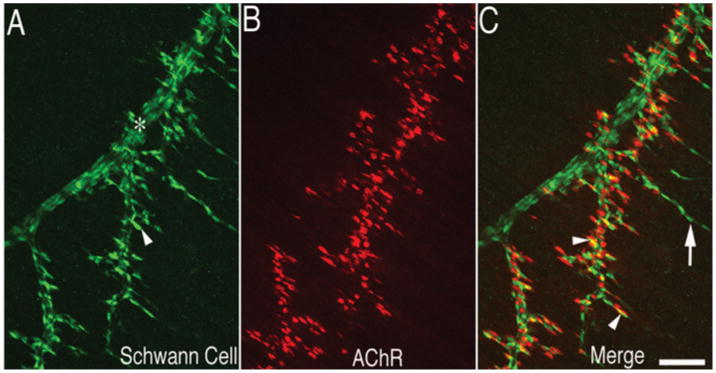
Figure 1. Distribution of Schwann cells along the nerve during neuromuscular synaptogenesis.

Mouse diaphragm muscle at E18.5 was immunolabelled with anti-S100 antibody (in green) for Schwann cells (A) and Texas Red-conjugated α-bungarotoxin (in red) for postsynaptic AChR (B). Schwann cells (arrowhead in A) delineanate both the nerve trunk (*) and the nerve branches and are present at the NMJ (arrowheads in C) as well as the tip of the nerve branches extended beyond the NMJ (arrow in C). Scale bar, 100 μm.
