
Figure 2. Ultrastructure of developing NMJs in embryonic muscles in mice.
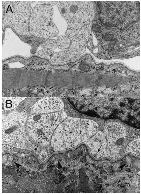
(A) A nerve–muscle contact depicting an early stage of synaptogenesis (E15.5 diaphragm). A cluster of axons loosely wrapped by immature Schwann cell attaches on to the muscle membrane. Although basal lamina is clearly seen at the synaptic cleft, presynaptic and postsynaptic specialization are barely recognizable. Only a few synaptic vesicles are present in the nerve terminal. (B) An immature NMJ (E18.5 diaphragm) illustrating primitive junctional folds (arrowheads) and organelles in the subsynaptic salcoplasm. Nerve terminals contain more synaptic vesicles compared with earlier stage shown in (A). Some synaptic vesicles (arrow) are clustered at the presynaptic membrane, presumably the active zone. Scale bar, 1 μm.