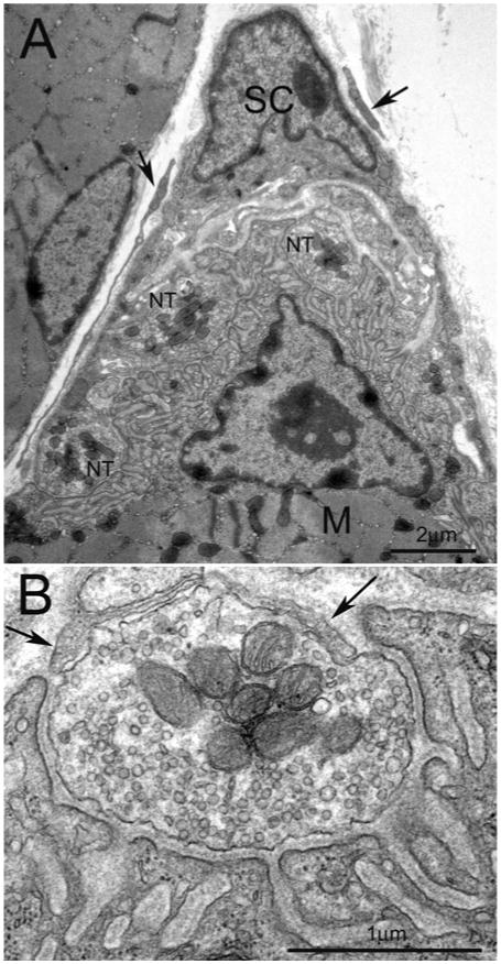
Figure 3. Ultrastructure of a mature NMJ in adult muscle in mice.

(A) Low magnification of electron micrograph illustrating typical arrangement of the NMJ. The Schwann cell (SC) with its electro-dense nucleus cap the nerve terminals (NT) apposing the postsynaptic specialization of the muscle (M). The processes (arrows) around Schwann cell may be the NMJ-capping cells recently characterized by Court et al. [12]. (B) Higher magnification of one of the nerve terminals shown in (A). The nerve terminal, half buried in the surface of muscle fibre, contains plentiful synaptic vesicles and mitochondria. Postsynaptic muscle membrane displays fully elaborated junctional folds. Basal lamina appears in the synaptic cleft. Opposing the muscle, thin processes of Schwann cell (arrows) cap the nerve terminal. Scale bar, 2 μm in (A) and 1 μm in (B).
