
Fig. 1.
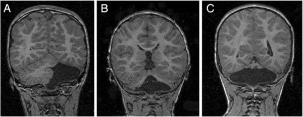
Follow-up brain MRIs (coronal spoiled gradient recalled T1-weighted) of infants with isolated cerebellar hemorrhagic injury on neonatal cranial ultrasound. a Complete absence of the left cerebellar hemisphere with preservation of the right cerebellar hemisphere and vermis. b Absence of the inferior cerebellar vermis and inferior portions of both cerebellar hemispheres. c Near-total cerebellar destruction with only a small amount of superior cerebellar vermis present (Reprint from [22] Copyright 2001 by American Academy of Pediatrics. Reprint with permission.)