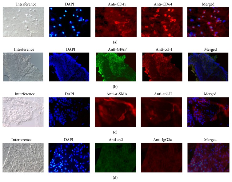
Figure 2.
Interference microscopy, cell nuclei staining with 4′,6-diamidino-2-phenylindole, DAPI (blue), and immunocytochemical staining of lamellar hole-associated epiretinal proliferation removed from eyes with lamellar macular holes (LMH). (a) Epiretinal cells show positive immunolabelling with anti-CD45 (red) and anti-CD64 (red) in specimen removed from eyes with LMH. (b) Immunostaining of epiretinal cells seen as a thick homogenous layer positively labelled with anti-GFAP (green) and anticollagen type I (anti-col-I) (red). (c) Immunolabelling with anti-α-smooth muscle actin (α-SMA) (red) and anticollagen type II (anti-col-II) (red). (d) Negative control specimen with positive cell nuclei staining but no specific immunoreactivity of cell proliferation. (Original magnification: (a) ×400; (b) ×100; (c-d) ×400).

