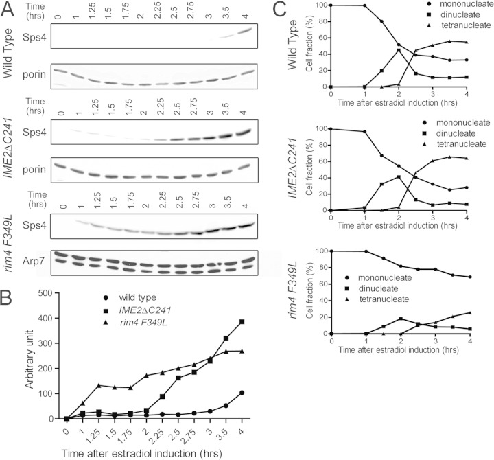
FIG 2.
Timing of Sps4 translation in wild-type and IME2ΔC241 cells. (A) The levels of Sps4-3HA (strains yLJ80 and yLJ89) or Sps4-GFP (strain yLJ167) were monitored by Western blotting. Cells were incubated in SPM for 6 h prior to the addition of β-estradiol (time zero), and aliquots were removed at the indicated times after induction. As a loading control, the same samples were probed with antibodies to the mitochondrial Por1 protein. (B) Quantitation of the levels of Sps4 in panel A. The values are normalized to the amount of Por1 protein in each lane for yLJ80 and yLJ89 and the amount of Arp7 protein for yLJ167. (C) DAPI (4′,6-diamidino-2-phenylindole) staining was performed to monitor the progression of cells through the meiotic divisions in the same time courses. The percentages of cells containing one nucleus (before MI), two nuclei (MI), or four nuclei (MII or later) are indicated.

