
FIG 1.
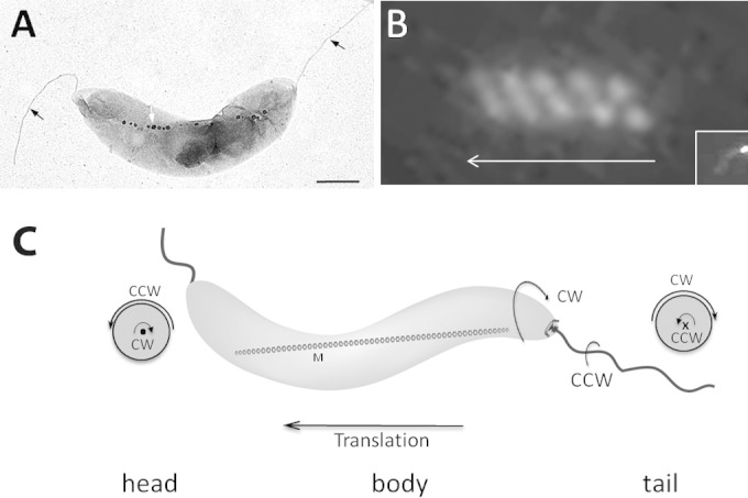
Rotation direction of AMB-1 cell body during runs. (A) Electron micrograph of a spirillum-shaped AMB-1 cell showing its two polar flagella (black arrows) and its magnetosome chain (white arrow). Bar, 500 nm. (B) Swimming trajectory reflected by single polar fluorescence labeling (inset) of MamU-GFP recorded during cell translation with a 100-ms exposure. The arrow indicates the direction of cell movement. (C) Schematic representation of an AMB-1 cell showing cell body and flagellar rotation directions. • and × show views of the flagella out-off and into the image, respectively.