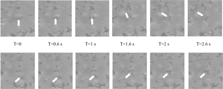
FIG 2.
Clumping in A. brasilense. The image sequences represent frames taken at 0.6-s intervals (T, time) from a video recording of free-swimming A. brasilense cells. The cultures were prepared by growing A. brasilense from a single colony, under elevated aeration, as described previously (8). The images were obtained by dark-field microscopy at ×40 magnification. Cells in transient stable clumps are visible. The arrows point to instances when a motile cell leaves a clump (top row) or joins a clump (bottom row). Cells in the suspension and the clumps were motile.

