
Fig. 9.
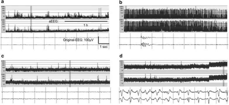
A simplified electroencephalogram (EEG) with two original EEG curves (lower panel), in combination with an amplitude integrated EEG (aEEG) trend curve (upper panel). Four dominating EEG patterns after cardiac arrest are shown: a flat, b suppression-burst, c continuous and d electrographic status epilepticus. From Friberg et al. [161]. Reprinted with permission