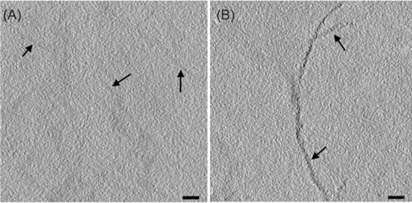
Fig. 1.

Section labelling of wild-type (A, arrows) and MTH-labelled (B, arrows) desmin. Samples prepared as described (Table S1, condition #10) were treated first with aurothiomalate, then a gold-enhancing solution Nanoprobes™ GoldEnhance EM. They were imaged without additional heavy metal staining. The labelling brought on by the attached MTH is evident in these 5-nm-thick tomographic slices. Bars = 50 nm. The distribution of label for this and other figures in the paper is more clearly seen in movies that show the 3D structures with serial computed sections. See Supplementary Materials.
