Figure 2.
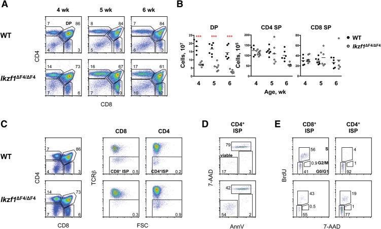
T-cell differentiation in Ikzf1ΔF4/ΔF4 mice progresses through a CD4+ ISP stage instead of a CD8+ ISP stage. (A) Shown are staining profiles of thymocytes in wild-type (WT) and Ikzf1ΔF4/ΔF4 mice at 4, 5, and 6 wk of age. The absolute mean percentages are indicated (n = 5–8). (B) Shown are absolute numbers of the indicated thymocyte populations in wild-type and Ikzf1ΔF4/ΔF4 mice at 4 wk (n = 5–8), 5 wk (n = 6–7), and 6 wk (n = 5–10). SP cells are gated on TCRβhi. (C) Shown are staining profiles of total (left), CD8+ gated (middle), and CD4+ gated (right) thymocytes of 4-wk-old wild-type and Ikzf1ΔF4/ΔF4 mice. The absolute percentages indicated are representative of five to eight mice. (D) Shown are staining profiles of Annexin V (AnnV) and 7-AAD (DNA content) for wild-type and Ikzf1ΔF4/ΔF4 TCRβlo CD4+ ISP thymocytes; the fractions of AnnV− 7-AAD− viable cells, AnnV+ 7-AAD− early apoptotic cells, and AnnV+ 7-AAD+ dead cells are indicated. Data are representative of three independent experiments. (E) Shown are cell cycle analyses of TCRβlo CD8+ gated (left) and TCRβlo CD4+ gated (right) cells using BrdU and 7-AAD (DNA content) staining, with cells in the G0/G1, S, and G2/M phases gated. Percentages are representative of three independent experiments.

