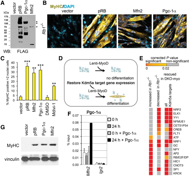
Figure 6.
Increased mitochondrial function rescues differentiation. (A) Immunoblot analysis of lysates prepared from cells expressing Flag-tagged proteins pRB, Mfn2, and Pgc-1α. (B) Differentiation rescue with mitochondrial regulators. Images of ICC for MyHC were generated using confocal microscopy. Rb1−/− MEFs containing MyoDER[T] lentiviruses were transduced with lentiviral vectors expressing pRB, Mfn2, and Pgc-1α proteins as in A or with the empty vector and induced with OHT. (C) Cells with increased mitochondrial function show high MyHC expression. MEFs containing MyoDER[T] were transduced with lentiviruses expressing the mitochondrial regulators or treated with 30 µM Drp1 inhibitor Mdivi-1. Empty vector or DMSO treatment was used as a control. Cells were analyzed after a 72-h treatment with OHT. Mean ± SD for the number of MyHC-positive cells with more than two nuclei in n = 5 microscopic fields (∼100 nuclei per field) in three representative experiments; (**)P < 0.001; (***) P < 0.0001, relative to the vector. (D) Schematics of myogenic differentiation rescue. Rb1−/− cells carrying Lenti-MyoD (or MyoDER[T]) were transduced with lentiviruses that express the mitochondrial regulators, restoring the expression level of Kdm5a targets. (E) Occurrences of TF-binding sites (TFBSs) in the promoter regions of Kdm5a targets. Enriched TFBSs with corrected P-values of <10−16 are shown in the heat map, arranged in the order of significance for genes increased in Rb1−/− and for the Kdm5a target genes rescued in DKO-myo. The whole list of enriched TFBSs with P < 0.02 is shown in Supplemental Table 4. (F) Pgc-1α specifically binds to the Mfn2 promoter during differentiation. ChIP experiments in Rb1−/− cells that were transduced with Pgc-1α lentiviruses and analyzed at 0 h and 24 h after induction. Igr2 is an intergenic control region. Mean ± SEM for n = 2 ChIP assays. (G) Expression level of MyHC is fully rescued by mitochondrial regulators. Immunoblot analysis of cell lysates prepared from Rb1−/− MEFs transduced with Lenti-MyoD and lentiviral vectors expressing pRB, Mfn2, or Pgc1α and induced for 120 h.

