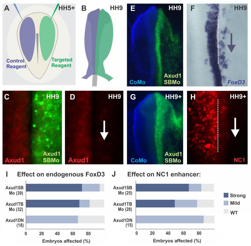
Figure 2. Axud1 is necessary for FoxD3 expression.
(A-B) Diagram of electroporation assays for loss-of-function experiments. (A) Control (blue) and targeted (green) reagents were injected in different sides of gastrulating embryos. (B) Following injection, embryos were electroporated and cultured until the desired stages. Phenotypes were scored by comparing the control and experimental sides of the same embryo. (C-D) Immunohistochemistry with an Axud1 antibody shows loss of the Axud1 protein in dorsal neural folds electroporated with Axud1 morpholino. (E-H) Morpholino-mediated knockdown of Axud1 resulted in a marked reduction of endogenous FoxD3 expression (E-F), as well as loss in activity of the NC1 enhancer, which controls cranial expression of FoxD3(Simoes-Costa et al., 2012) (G-H). (I-J) Quantitation of embryos following loss-of-function assays. The total number of embryos analyzed for each experiment is represented in parenthesis. See Fig. S1 for additional controls of loss of function experiments. CoMo: Control morpholino, Axud1DN: Axud1 dominant negative, Axud1SBMo: Axud1 splice-blocking morpholino, Axud1TBMo: Axud1 translation-blocking morpholino, HH: Hamburger and Hamilton developmental stages.

