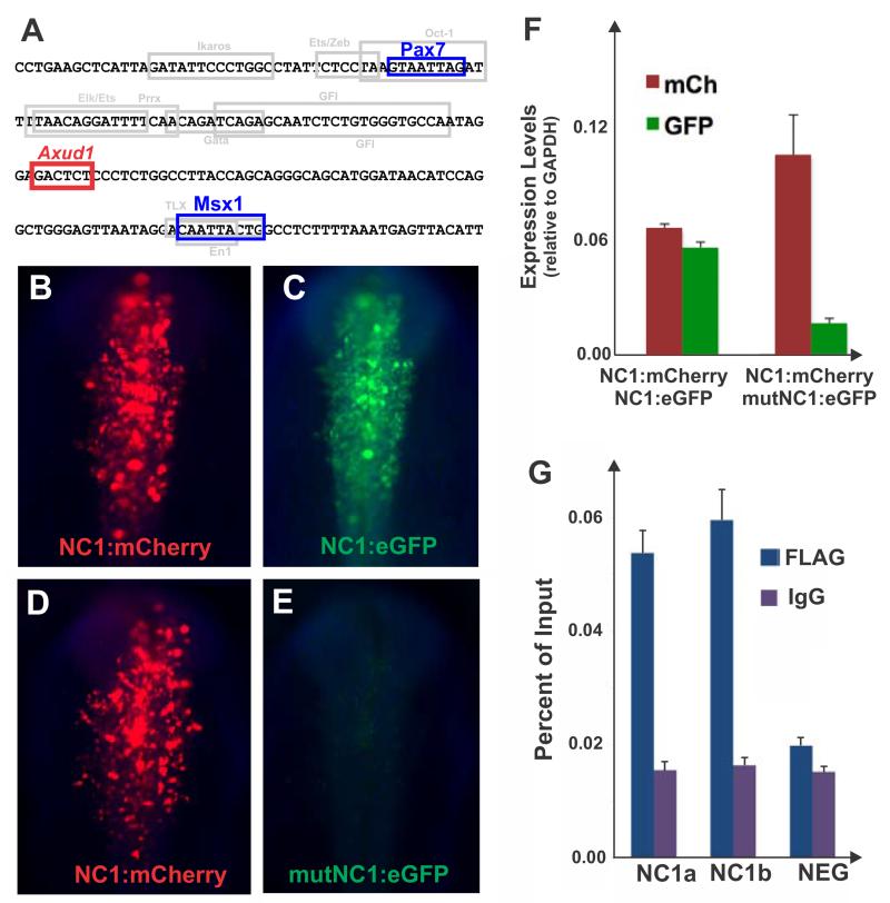
Figure 3. Axud1 is a direct regulator of neural crest specifier gene FoxD3.
(A) The NC1 enhancer core contains an Axud1 binding site between Pax7 and Msx1 sites. (B,C) Co-electroporation of a NC1 enhancer driving mCherry (NC1:mCherry) and an NC1 enhancer encoding eGFP (NC1:eGFP) result in similar levels of reporter gene expression in the same embryo. (D, E) Alternatively, co-electroporation of a NC1:mCherry with a mutated Axud-binding site encoding GFP (mut:NC1:eGFP) indicate the construct harboring the mutation (E) has negligible activity when compared to the wild type mCherry vector (D). (F) Quantitation of reporter gene expression levels through qPCR confirmed mutation of the Axud1 binding site results in loss of NC1 enhancer activity. Reporter gene levels are represented relative to GAPDH expression. (G) Chromatin immunoprecipitation assay performed in dissected dorsal neural folds of stage HH9 embryos electroporated with a FLAG-tagged Axud1 expression construct. Immunoprecipitation of the Axud1 protein with an anti-FLAG antibody, followed by site specific qPCR with primer pairs designed to amplify two fragments within the NC1 region (NC1a and NC1b) revealed significant enrichment of the NC1 region in the Axud1 pull-down when compared to IGG controls. No enrichment was observed for a negative control region upstream of the FoxD3 locus in the chicken chromosome 8 (NEG). Enrichment of amplicons is expressed as a percent of the total input chromatin used in the immunoprecipitation assay. Error bars on F and G represent standard deviation.

