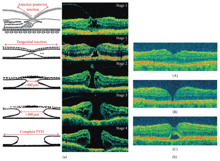
Figure 2.
(a) Schematic and OCT representation of macular hole formation [1]. (b) Optical coherence tomography images 3 months after macular hole surgery. (A) Normal gross anatomic features with an attached fovea. (B) Flat edges with persistent neurosensory defect. (C) Contiguous foveal surface with persistent subfoveal fluid [1].

