
Fig. 4.
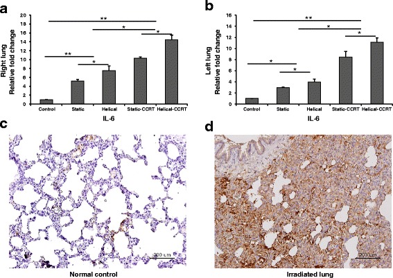
Tissue expression of IL-6. a Right lung samples demonstrated higher tissue IL-6 expression compared to the untreated control samples. b Left lung samples revealed upregulated tissue IL-6 expression. Immunohistochemical staining with anti-IL-6 antibody of c normal control and d right lung sample treated with helical tomotherapy with CCRT. * p <0.05; ** p <0.01; IL, interleukin; CCRT, concurrent chemoradiotherapy; bars = 200 μm