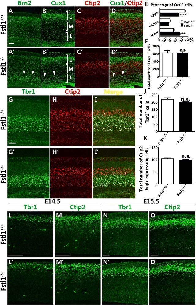
Fig. 2.

Disruption of Fstl1 results in an abnormal distribution of upper-layer but not deeper-layer cortical neurons. a to d’ The abnormal distribution of upper-layer neurons in the Fstl1 −/− cerebral cortex. Immunostaining for Brn2 (arrowheads, [a’]) and Cux1 (arrowhead, [b’]) at E18.5 shows many upper-layer neurons still residing in the IZ and subplate after Fstl1 ablation (arrowhead in a’ and b’). Co-immunostaining for Cux1 (b, b’, d and d’) and Ctip2 (c, c’, d and d’) at E18.5 shows the abnormal distribution of upper-layer neurons in the deeper area. e The distribution of Cux1+ neurons at E18.5. More Cux1+ neurons accumulated in the lower area at E18.5 (24.31 ± 1.26 % for WT, n = 4; 35.94 ± 0.48 % for Fstl1 −/−, n = 4, **p < 0.01), while fewer Cux1+ neurons were present in the upper area (44.14 ± 1.03 % for WT, n = 4; 32.98 ± 1.16 % for Fstl1 −/−, n = 4, ***p < 0.001). f The total number of Cux1+ neurons per area at E18.5. The data are the mean ± s.e.m.: 623.5 ± 58.5 for WT, n = 4; and 622.3 ± 29.1 for Fstl1 −/−, n = 4, p = 0.99. g to i’ Double immunostaining for Tbr1 and Ctip2 at E18.5 showed that the distributions of layer VI and layer Va neurons are unchanged in the Fstl1 −/− cortex compared to the WT cortex. j and k The total numbers of Tbr1+ layer VI neurons (j) and layer Va neurons with high Ctip2 expression per area at E18.5 (k). The Tbr1+ neuron data are presented as the mean ± s.e.m.: 220.5 ± 8.4 for WT, n = 4; and 198.4 ± 4.7 for Fstl1 −/−, n = 4, p = 0.07. The high Ctip2 expression neuron data are presented as the mean ± s.e.m.: 104.5 ± 3.0 for WT, n = 5; and 99.4 ± 2.6 for Fstl1 −/−, n = 5, p = 0.24. l to o’ The comparable distribution of deeper-layer neurons at early embryonic stages in WT and Fstl1 −/− mice. Immunostaining for the deeper-layer neuronal markers Tbr1 (l, l’, n and n’) and Ctip2 (m, m’, o and o’) at E14.5 (l to m’) and E15.5 (n to o’) shows that the migration and distribution of deeper-layer neurons are unchanged in the Fstl1 −/− cortex compared to the WT cortex. Scale bars: 100 μm
