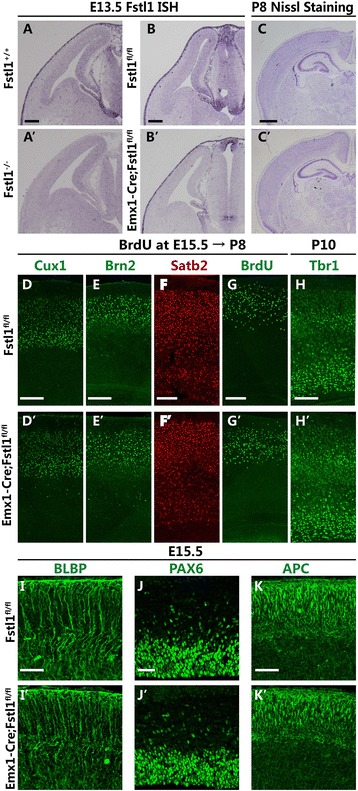
Fig. 8.

Fstl1 deletion in the VZ only was not sufficient to induce radial glial dysplasia. a to b’ A high level of Fstl1 mRNA can be detected in the pia mater and VZ in the WT (a) and Fstl1 fl/fl (b) cortices. In the Fstl1 −/− mice, Fstl1 mRNA cannot be detected in the pia mater or the cerebral cortex (a’); in the Emx1 IREScre; Fstl1 fl/fl mice, Fstl1 expression was absent in the VZ but present in the pia mater (b’). c and c’ Nissl staining showed no remarkable differences in the structure or cellular distribution of the cerebral cortex between the Fstl1 fl/fl (c) and Emx1 IREScre ; Fstl1 fl/fl (c’) mice at P8. d to h’ Immunolabelling with anti-Cux1 (d and d’), anti-Brn2 (e and e’) and anti-Satb2 (f and f’) antibodies at P8 and with anti-Tbr1 (h and h’) antibodies at P10 showed that both the upper- (d to f’) and the deeper-layer (h and h’) cortical neurons were nicely laminated and eventually migrated to the appropriate cortical layers in the Emx1 IREScre; Fstl1 fl/fl mice (d, e, f and h), similar to observations in the Fstl1 fl/fl mice (d’, e’, f’ and h’). g and g’ There were no differences in the final destinations of the cells that were labelled with BrdU at E15 between the Emx1 IREScre; Fstl1 fl/fl and Fstl1 fl/fl cortices at P8. i and i’ BLBP immunostaining showed normal radial glial development in the Fstl1 fl/fl (i) and Emx1 IREScre; Fstl1 fl/fl cortices (i’) at E15.5. j and j’ Pax6 immunostaining showed no obvious differences between the E15.5 Fstl1 fl/fl and Emx1 IREScre; Fstl1 fl/fl cerebral cortices. k and k’ Normal basal branching of the radial glial, as demonstrated by immunolabelling for APC in the Emx1 IREScre; Fstl1 fl/fl (k’) and Fstl1 fl/fl (k) cortices. Scale bars: 100 μm for a–b’; 200 μm for c to h’; 50 μm for i–k’
