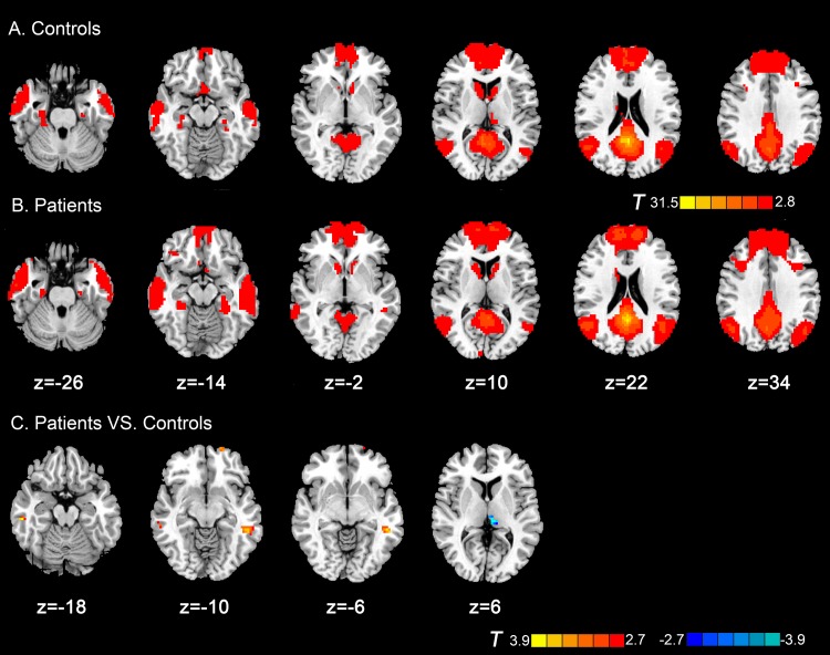
Fig 1. Axial magnetic resonance (MR) images show resting-state functional connectivity (rsFC) with posterior cingulate cortex within and between groups.
Regions show significantly positive functional connectivity in (A) control subjects and (B) patients with subcortical sVCI (p < 0.05, AlphaSim corrected). (C) Compared with the control group, patients with sVCI exhibited increased rsFC in the left middle temporal lobe, right inferior temporal lobe and left superior frontal gyrus. The left thalamus exhibited decreased connectivity (p < 0.05, AlphaSim corrected). The t-score bars are shown on the right. Red indicates patients with sVCI > control and blue indicates patients with sVCI < control. Note: The left part of the figure represents the participant’s left side, the right part represents the participant’s right side. sVCI = subcortical vascular cognitive impairment.

