Fig. 1.

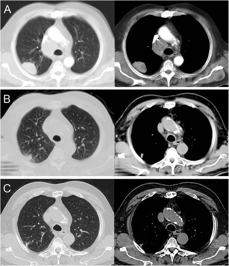
a Initial chest computed tomography scan showing the tumor in the upper lobe of the right lung and enlarged mediastinal lymph nodes in the paratracheal position. b Computed tomography scan after 2 months, c after 1 year

a Initial chest computed tomography scan showing the tumor in the upper lobe of the right lung and enlarged mediastinal lymph nodes in the paratracheal position. b Computed tomography scan after 2 months, c after 1 year