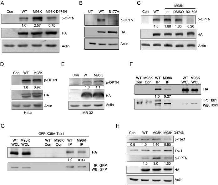
Fig 3. M98K-OPTN shows enhanced Tbk1 dependent phosphorylation.
(A) Western blots show p-OPTN (p-S177) levels in RGC-5 cells infected with control (con), WT-OPTN, M98K-OPTN or M98K-D474N-OPTN adenoviruses after 24 hrs of expression. HA blot shows expression levels of various constructs of OPTN. Actin was used as a loading control. The numbers below p-OPTN blot indicate relative p-OPTN levels after normalization with OPTN expression blot. (B) Western blots show p-OPTN (Ser177) levels in RGC-5 cells transfected with WT-OPTN or S177A-OPTN expression vector after 24 hrs of expression. UT, untransfected. (C) Inhibition of Tbk1 reduces M98K-OPTN phosphorylation. RGC-5 cells were infected with control (con), WT-OPTN or M98K-OPTN adenoviruses; M98K adenovirus infected cells were either kept untreated (ut) or treated with DMSO or with 1 μM BX-795 for 18 hrs. Cell lysates were then subjected to western blotting. (D and E) Expression of M98K-OPTN in HeLa or IMR-32 cells does not result in enhanced phosphorylation. Relative p-OPTN levels in Hela and IMR-32 cells respectively, after control (con) WT-OPTN or M98K-OPTN adenovirus infection. (F) Interaction of OPTN with Tbk1 in retinal cells. RGC-5 cells were infected with adenoviruses expressing HA-tagged WT-OPTN or M98K-OPTN for 24 hrs and cell lysates were subjected to immunoprecipitation with Tbk1 antibody or control antibody (Con) and western blotting done with HA and Tbk1 antibodies. WCL, whole cell lysate. The numbers below HA blot indicate relative levels of co-purified WT-OPTN and M98K-OPTN levels after normalization with immunoprecipitated Tbk1 protein. (G) Interaction of catalytically inactive mutant of Tbk1, K38A-Tbk1 with WT-OPTN and M98K-OPTN in retinal cells. RGC-5 cells were co-transfected with plasmids expressing HA-tagged WT-OPTN or M98K-OPTN along with GFP-tagged K38A-Tbk1 for 24 hrs and cell lysates were subjected to immunoprecipitation with GFP antibody or control antibody (Con) and western blotting done with HA and GFP antibodies. WCL, whole cell lysate. The numbers below HA blot indicate relative levels of co-purified WT-OPTN and M98K-OPTN levels after normalization with immunoprecipitated K38A-Tbk1 protein. (H) M98K-OPTN activates Tbk1. Western blots show p-Tbk1 (Ser172) levels in RGC-5 cells infected with control (con), WT-OPTN, M98K-OPTN or M98K-D474N-OPTN adenoviruses after 24 hrs of expression. HA blot shows expression levels of various constructs of OPTN. Actin was used as a loading control. The numbers below p-Tbk1 blot indicate relative p-Tbk1 levels after normalization with Tbk1 blot. The number below p-OPTN blot indicates relative p-OPTN levels after normalization with expression blot.

