. 2015 Sep 9;45(3):187–192. doi: 10.5624/isd.2015.45.3.187

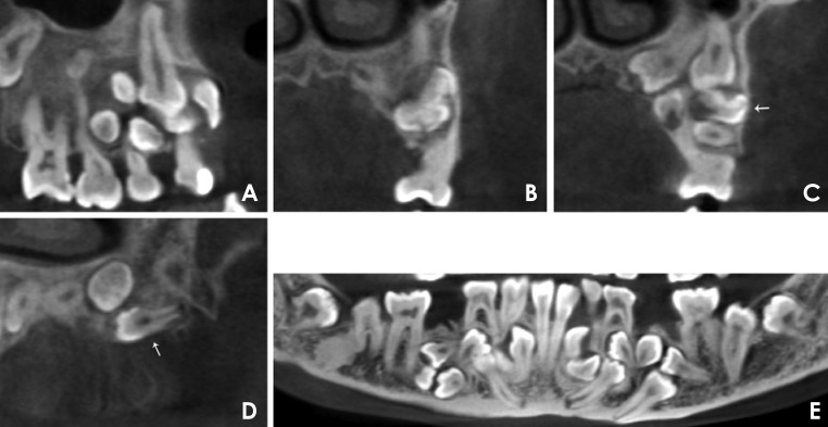
Fig. 7. A. A panoramic reformatted cone-beam computed tomography (CBCT) image shows a 24.8 mm×14.0 mm radiolucency with a well-defined margin in the right premolar region of the maxilla. B. A coronal CBCT image shows an odontoma. The odontoma in the left premolar region of the maxilla presented as a 12.8 mm×6.9 mm irregular radiopacity surrounded by a radiolucent area. C. A coronal CBCT image shows part of the odontoma (arrow) and four supernumerary teeth in the left maxilla. The supernumerary teeth are different in terms of direction (normal, buccolingual, or inverted) and root development. D. A sagittal CBCT image shows a supernumerary tooth (arrow) that grew from the distal to mesial position in the left maxilla. E. A panoramic reformatted CBCT image shows fusion between the right mandibular deciduous lateral incisor and canine, which seems to be incomplete, sharing part of the pulp chamber with separate root canals. The trabeculation in the mandible is coarse and disordered.

Fig. 7