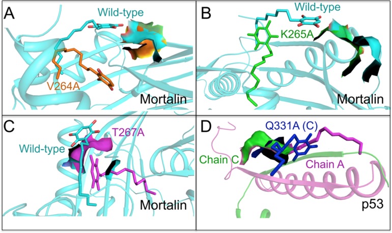
Fig 5. Molecular docking analysis of embelin with mutant mortalin and p53 proteins.
The change in conformation of embelin and binding site in (A) wild type (light blue) and V264A (orange) mortalin (B) wild type (light blue) and K265A (green) mortalin (C) wild-type (light blue) and T267A (purple) mortalin and (D) wild type chain C (green) and Q331A chain C (blue) p53.

